


Content of the article:
X-ray examination
An integral part of the examination in the dental clinic is an X-ray examination, based on which it is possible to properly evaluate and diagnose all the problems that are present in your dentition. These are often hidden problems that are not visible to the naked eye.
There are several types of dental radiographs. We divide them into extra-oral (external), when the X-ray film is outside the patient’s mouth, and intra-oral (internal), when the film is inserted into the mouth.
Extraoral radiographs
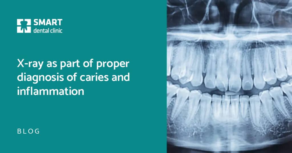
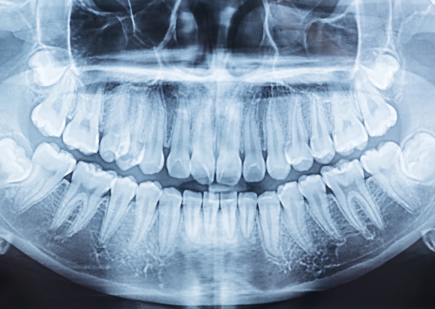
Orthopantomography (OPG)
A panoramic image that shows the entire dentition, i.e., the upper and lower jaw, as well as adjacent anatomical structures. It gives us an overall view of your teeth, thanks to which we can diagnose or rule out, for example, inflammations of the bone around the teeth, bone retreat, parodontitis, tooth fractures or cystic formations.
We can also monitor the condition of the sinuses, temporomandibular joints, the position of teeth in the dental arches, the position of wisdom teeth or the establishment and eruption of milk teeth in children. OPG is appropriate at initial patient examination and is recommended to be repeated every 2 years. Based on it, more detailed images may subsequently be required.
Intraoral radiographs
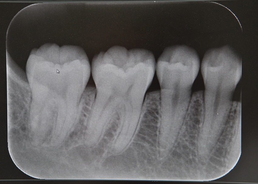
Intraoral periapical image
It shows in detail the entire tooth, that is, the crown part but also its root part, which is essential for the diagnosis of inflammation in the root tip area and painful conditions with which the patient comes to the dental clinic. This examination is also important in the endodontic treatment of root canals, when we check whether the canals have been filled with the root filler correctly and evenly all the way to their end, or we can check the quality of the previous root filler.
BTW, the so-called bite snapshot
It serves the purpose of a more detailed diagnosis of the crown part of individual teeth, thanks to which we are able to detect mainly hidden cavities that may be found in the interdental spaces and under old fillings.
This X-ray will also show the relationship of old fillings to the medullary cavity, and we can also diagnose secondary caries that may sooner or later pose a risk to the dental marrow, cause toothache and, consequently, death of the dental nerve.
The sooner we expose these hidden caries, the more likely the tooth can be saved. Bite snapshots should also be taken as part of the initial examination and at least once a year during the preventive check-up.
Utilization of x-ray imaging
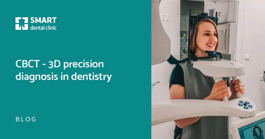
X-ray examinations are used for diagnosis and for subsequent design of treatment plan to be carried out in our clinic, all in one place. We have state-of-the-art X-ray instruments and all images are taken in digital form (radiography), which allows us to see them immediately in the office on the computer screen. In addition, the radiation exposure is significantly reduced. We use protective devices during the examination.
DID THE ARTICLE RESONATE WITH YOU?
BOOK AN EXAMINATION TODAY!
-
X-ray images from 12 - 28 €
of the bite, detailed, an OPG image
-
CT scan from 65 - 95 €

MDDr. Ján Šimon
Dentist, Bratislava