X-ray examination
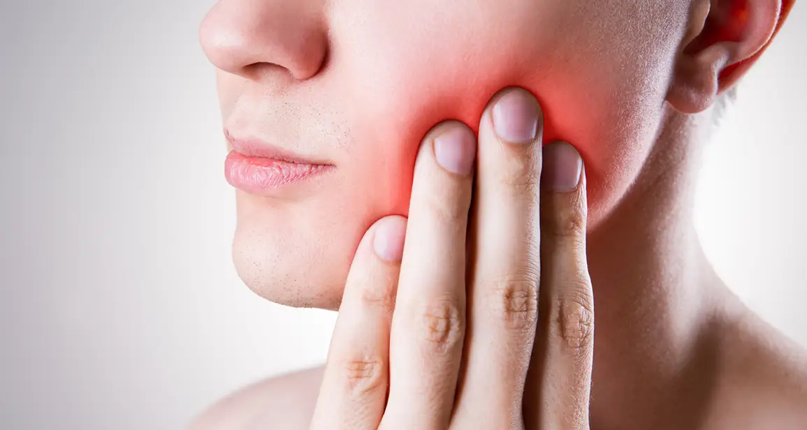
An indispensable part of a dental examination is an X-ray, which allows for the correct evaluation and diagnosis of all problems present in your dentition. These are often hidden problems that are not visible to the naked eye.
There are several types of dental X-ray images. They are divided into extraoral (external), where the X-ray film is located outside the patient's mouth, and intraoral (internal), where the film is placed inside the mouth.
Extraoral X-ray images
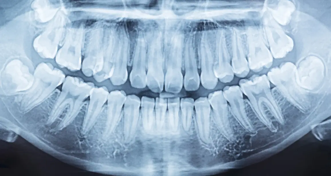
Orthopantomographic image (OPG)
A panoramic image that displays the entire dentition, including both the upper and lower jaws, as well as adjacent anatomical structures. It provides a comprehensive view of your dentition, allowing us to diagnose or rule out, for example, bone inflammation around the teeth, bone loss, periodontitis, tooth fractures, or cystic formations.
We can also monitor the condition of the paranasal sinuses, temporomandibular joints, the position of teeth in the dental arches, the position of wisdom teeth, or the development and eruption of deciduous teeth in children. An OPG is suitable for a patient's initial examination and is recommended every 2 years. Based on this, more detailed images can then be indicated.

Intraoral X-ray images
Intraoral periapical image
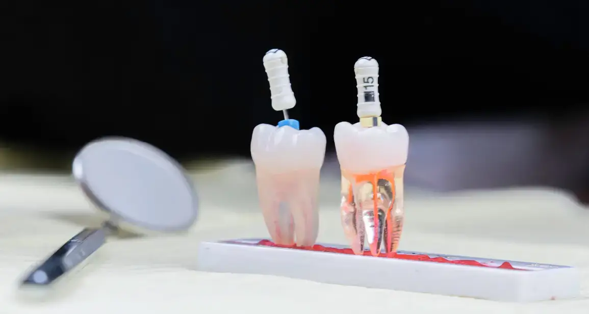
It shows the entire tooth in detail, meaning both its crown and root parts, which is essential for diagnosing inflammation at the root apex and for painful conditions that bring the patient to the dental office. This examination is also important during endodontic treatment of root canals, when we check whether the canals have been filled correctly and evenly with the root filling all the way to their end, or we can check the quality of a previous root filling.

Bitewing X-ray (BTW), also known as a bite-wing image
It serves for more detailed diagnosis of the crown part of individual teeth, allowing us to detect hidden cavities, especially those located in interdental spaces and under old fillings. This X-ray also shows the relationship of old fillings to the pulp cavity, and we can also diagnose secondary cavities which, sooner or later, can threaten the dental pulp, cause tooth pain, and eventually lead to the death of the dental nerve. The sooner these hidden cavities are detected, the greater the chance of saving the tooth. Bitewing X-rays are also suitable for initial examinations and at least once a year during preventive check-ups.
the newsletter
By submitting this form, you consent to the processing of your email address and/or phone number for the purpose of sending commercial and non-commercial messages.
Use of X-ray images
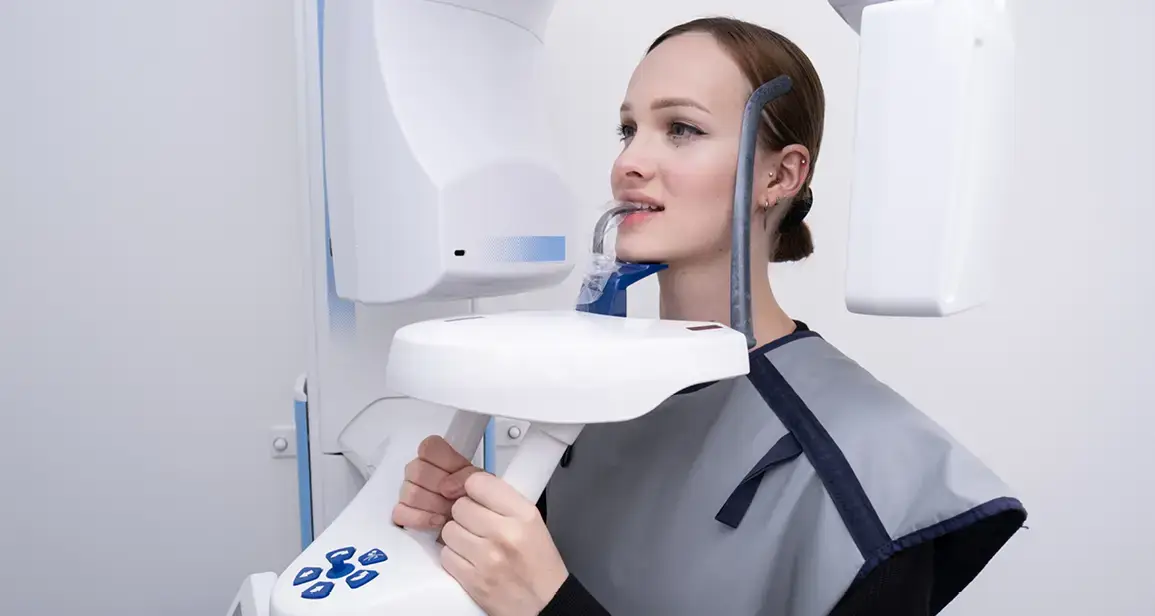
We use X-ray examinations for diagnosis and for the subsequent creation of a treatment plan right here at our clinic, all in one place. We have modern X-ray equipment available, and all images are taken digitally (radiovisiography), which allows us to view them immediately on the monitor in the office and also significantly reduces the radiation dose. We use protective measures during the examination.


Dentist
I completed my dental medicine studies in 2020 at the Faculty of Medicine, Comenius University in Bratislava. During my studies, I completed several internships at modern clinics in Slovakia and participated in… Read more